The Minnesota Starvation Experiment, conducted between November 1944 and December 1945 at the University of Minnesota, remains one of the most rigorous clinical investigations into human energy deprivation, metabolic adaptation, and refeeding recovery. Led by Dr. Ancel Keys (a sociopath who had no qualms about unethical activity) and published in 1950 as the comprehensive two-volume treatise The Biology of Human Starvation, this study mapped the physiological, endocrinological, and psychological boundaries of severe caloric restriction.
While initially commissioned by the U.S. Office of the Surgeon General to formulate scientific protocols for post-WWII famine relief in Europe, its findings provide profound baseline insights into metabolic rate regulation, body composition shifts, adaptive thermogenesis, and the severe neurobehavioral impacts of sustained energy deficits.
Study Design and Experimental Architecture
The trial evaluated 36 healthy, psychologically screened male conscientious objectors selected from a pool of over 400 volunteers. The 13-month protocol was divided into four tightly controlled phases:
-
Phase 1: Baseline Control (12 Weeks): Participants consumed an individualized diet averaging 3,200 kilocalories per day to establish nitrogen balance, stable body mass, and baseline cardiovascular, metabolic, and psychological metrics.
-
Phase 2: Semi-Starvation (24 Weeks): Caloric intake was slashed by approximately 50%, averaging 1,560 kilocalories per day. The diet mimicked the high-carbohydrate, low-protein, low-fat famine rations typical of war-torn Europe (potatoes, rutabagas, turnips, whole wheat bread, and macaroni). Caloric allotments were adjusted weekly for each subject to enforce a target 25% reduction in total body weight over 6 months, while requiring 22 miles of weekly walking and daily lab labor.
-
Phase 3: Controlled Rehabilitation (12 Weeks): The subjects were randomized into four refeeding cohorts receiving caloric increases of 400, 800, 1,200, or 1,600 kilocalories above the starvation baseline. Sub-groups were tested with protein and vitamin supplementation to identify the exact nutritional thresholds required for tissue repair.
-
Phase 4: Unrestricted Rehabilitation (8 Weeks): Subjects were permitted unrestricted ad-libitum intake (at one’s pleasure” or “as desired”). Caloric consumption, body weight velocity, and metabolic restoration were continuously tracked without dietary constraints.
Metabolic and Endocrinological Adaptations
Prolonged energy deficiency triggered intense biological survival mechanisms aimed at minimizing energy expenditure:
-
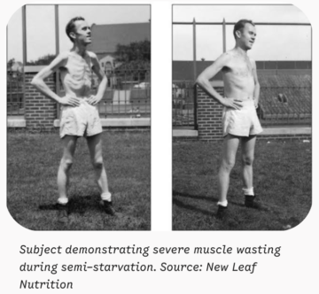
Basal Metabolic Rate Reduction: Basal Metabolic Rate (BMR) dropped by approximately 40% from baseline. Detailed tissue analysis demonstrated that only roughly half of this reduction was attributable to the physical loss of active cell mass (lean tissue organ and muscle mass). The remaining half resulted from true “adaptive thermogenesis”—a profound downregulation of cellular metabolic rate per unit of living tissue.
-
Body Composition Alterations: Subjects lost an average of 25% of overall body weight. Tissue loss was non-uniform: fat mass was depleted by ~70%, while total lean body mass fell by ~40%.
-
Cardiovascular Collapse and Autonomic Shifts: Resting heart rates plummeted from a pre-study mean of 55 beats per minute down to as low as 35 beats per minute. Cardiac muscle mass decreased by ~20%, stroke volume dropped substantially, blood pressure declined, and total circulating blood volume contracted by approximately 10%.
-
Famine Edema: Despite normal plasma protein levels, subjects developed clinical extracellular fluid retention (famine edema), manifesting as soft tissue swelling in the knees, ankles, and face.
-
Somatic and Hormonal Suppression: Core body temperature dropped by up to 1 degree Fahrenheit, resulting in severe cold intolerance. Thyroid activity downregulated, gastrointestinal motility decreased dramatically (with bowel movements slowing to once per week for some subjects), and hair growth slowed significantly.
Cellular Mechanisms Behind Adaptive Thermogenesis “Deep Dive”
WARNING: This section is for science geeks like myself. If this isn’t you, skip down to the next section, “Neuropsychiatric and Behavioral Impact”
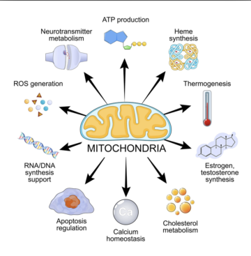
Adaptive thermogenesis refers to a regulated decrease in energy expenditure beyond what changes in lean and fat body mass can explain. At the cellular level, the human body executes a coordinated metabolic shutdown across mitochondrial, enzymatic, and neuroendocrine systems to preserve adenosine triphosphate (ATP) supplies necessary for survival.
1. Mitochondrial Coupling Efficiency and Proton Leak Suppression
In energy-abundant conditions, a significant portion of basal metabolic rate (~20-30%) is expended through “basal proton leak” across the inner mitochondrial membrane. Protons pumped into the intermembrane space by the electron transport chain (Complexes I, III, and IV) leak back into the mitochondrial matrix independently of ATP synthase via uncoupling proteins (UCP1 in brown/beige adipose tissue, and UCP2/UCP3 in skeletal muscle and cardiac tissue). This process dissipates the proton motive force as thermal energy (heat) rather than capturing it as cellular energy (ATP).
During sustained starvation, the cell downregulates the expression and activity of UCP2 and UCP3. By reducing basal proton leak, mitochondria become tightly coupled. Higher mitochondrial coupling efficiency means that more ATP is synthesized per mole of substrate oxidized, minimizing heat dissipation and drastically lowering substrate utilization at rest.
2. Thyroid Hormone Deiodination Dynamics
Thyroid hormones are primary regulators of nuclear transcription for metabolic machinery. In semi-starvation:
-
Peripheral Conversion Suppression: Expression of 5′-deiodinase enzymes (D1 and D2) decreases, reducing the peripheral conversion of inactive thyroxine (T4) into active triiodothyronine (T3).
-
Reverse T3 Accumulation: Alternative deiodination via 3-deiodinase increases, converting T4 into metabolic-inactive reverse T3 (rT3), which competitively inhibits active T3 signaling at cellular receptor sites.
-
Transcriptional Downregulation: Lower intracellular T3 levels reduce the nuclear transcription of critical metabolic genes, including mitochondrial enzymes (e.g., cytochrome c oxidase), glycerol-3-phosphate dehydrogenase, and ion transporters.
3. AMP-Activated Protein Kinase (AMPK) Activation
As intracellular ATP stores decrease and the AMP/ATP ratio rises, 5′-AMP-activated protein kinase (AMPK) acts as a master energy sensor:
-
Inhibition of Anabolic Pathways: AMPK phosphorylates and inactivates acetyl-CoA carboxylase (ACC), shutting down de novo lipolysis and fatty acid synthesis. It simultaneously suppresses mammalian target of rapamycin complex 1 (mTORC1), terminating energy-expensive protein synthesis and cell growth.
-
Suppression of Futile Substrate Cycles: Cells downregulate energy-consuming “futile cycles” (such as the simultaneous operation of glycolysis and gluconeogenesis, or pyruvate-malate cycling), reducing ATP waste.
4. Sympathetic Tone and Adrenergic Signaling Downregulation
Extended caloric deficits cause a marked reduction in sympathetic nervous system (SNS) outflow from the brainstem:
-
Reduced Norepinephrine Release: Lower plasma norepinephrine concentrations lead to reduced occupancy of beta-1, beta-2, and beta-3 adrenergic receptors on cardiomyocytes, adipocytes, and skeletal muscle cells.
-
Attenuated cAMP/PKA Cascade: Lower adrenergic stimulation decreases intracellular cyclic AMP (cAMP) production and protein kinase A (PKA) activation. In adipose tissue, this suppresses hormone-sensitive lipase (HSL) activity to conserve lipid reserves; in thermogenic tissues, it removes the stimulus for brown adipose tissue activity.
5. Downregulation of Active Ion Transport Pumps
Maintaining transmembrane electrochemical gradients consumes approximately 25-30% of total resting cellular energy. Two major ATPases are target-suppressed during adaptive thermogenesis:
-
Sodium-Potassium Pump (Na+/K+-ATPase): Decreased T3 and adrenergic signaling directly reduce the transcription and membrane density of Na+/K+-ATPase pumps in skeletal muscle and visceral organs, lowering resting ATP consumption.
-
SERCA Pumps: Sarcoplasmic/endoplasmic reticulum calcium ATPase (SERCA) pumps, which transport Ca2+ ions back into the lumen of the sarcoplasmic reticulum during muscle relaxation, lower their turnover rate, decreasing skeletal muscle resting metabolic rate.
Neuropsychiatric and Behavioral Impact
The psychological shifts recorded during the 24-week semi-starvation phase highlighted the powerful connection between metabolic status and brain function:
-
Minnesota Multiphasic Personality Inventory (MMPI) Spikes: Serial testing revealed dramatic elevations in depression, hysteria, and hypochondriasis—a constellation clinical researchers termed the “neurotic triad.”
-
Obsessive Food Fixation: Subjects developed overwhelming cognitive fixations on nutrition. They spent hours reading cookbooks, collecting recipes, studying restaurant menus, and analyzing agricultural data. Mealtime behaviors turned obsessive, with subjects diluting food with water or adding excessive salt to artificially increase bulk, and chewing each bite endlessly to delay finishing.
-
Social and Affective Flattening: Libido dropped to zero. Apathy, irritability, and social isolation dominated. Voluntary physical activity ceased entirely outside mandatory walking requirements as subjects conserved every available calorie (click here for a deeper dive on the “supersaver” metabolism).
-
Refeeding Hyperphagia and Fat Overshoot: During the initial weeks of unrestricted rehabilitation, satiety signaling was entirely non-functional. Subjects routinely consumed 4,000 to 11,000 kilocalories per day. This period triggered “post-starvation fat overshoot,” a phenomenon where the body preferentially accumulated adipose tissue at a rate and total percentage far exceeding baseline levels before lean body mass could fully regenerate.
Key Takeaways for Human Physiology and Clinical Refeeding
The Minnesota Starvation Experiment established fundamental clinical principles that remain vital today:
-
Refeeding Requirements: The study demonstrated that low-calorie refeeding diets (under 3,000 kilocalories per day) failed to achieve physical recovery or reverse tissue wasting in starved individuals. Full recovery required sustained daily energy intakes of 4,000 kilocalories or more. Note: Since this experiment was conducted during World War II, most people’s metabolism then differed significantly from today’s because they were naturally more active and processed food was much less abundant (to name just a couple of the many reasons). I mention this because, due to modern humans’ lower metabolism, 4000 cal a day on the current Standard American Diet (SAD) would be too much, while it wasn’t for people back then.
-
Metabolic Flexibility Limits: Extreme, prolonged caloric deficits drive physiological compensations that suppress baseline energy expenditure far beyond what simple loss of muscle mass accounts for.
-
Tissue Regeneration Hierarchy: Following severe depletion, body fat mass recovers much faster than skeletal muscle and organ tissue, highlighting the challenge of restoring lean body mass without causing transient fat accumulation. In other words, this explains why consistent yo-yo dieting (the way most people do it nowadays) always results in a lower metabolism every time someone yo-yos.
Clinical Implications: Severe Caloric Restriction and Anorexia Nervosa
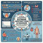
The profound metabolic adaptations demonstrated in the Minnesota Starvation Experiment mirror the pathophysiological mechanisms observed in clinical conditions of chronic energy deprivation, such as Anorexia Nervosa and extreme caloric restriction regimes. When energy intake remains chronically suppressed below basal requirements, the body enforces long-term structural and neuroendocrine adaptations to prioritize essential cellular survival over energetic maintenance. Sustained suppression of the hypothalamic-pituitary-thyroid (HPT) and hypothalamic-pituitary-gonadal (HPG) axes reduces circulating free T3, estrogen, and testosterone, while chronically elevating cortisol. This hormone profile drives severe loss of metabolically active organ parenchymal mass and skeletal muscle tissue. Concurrently, persistent reductions in leptin signaling and mitochondrial uncoupling protein expression reduce basal energy expenditure per unit of remaining lean tissue. This lingering biological state—termed persistent adaptive thermogenesis—can cause the body to burn significantly fewer calories than standard predictive metabolic models estimate, even months after increased energy intake. Consequently, individuals emerging from chronic starvation frequently encounter severe metabolic rigidity (aka “set point”), altered satiety signaling, and accelerated adipose accumulation relative to lean mass restoration (“post-starvation fat overshoot”), underscoring the necessity of structured, long-term physiological rehabilitation to rebuild active tissue mass (especially balanced macros and strength training) and restore a normal and healthy baseline metabolic rate.
References
Latest Posts